
|
|
Anup Shah
Resident, Dept. of Urology
UW School of Medicine
Marla Paun,
Engineer, CIMU
John Kucewicz,
Engineer, CIMU
Oleg Sapozhnikov,
Engineer, CIMU
Manjiri Dighe
Dept. of Urology
UW School of Medicine
Hunter McKay
Urologist
Seattle Polyclinic
Mathew Sorensen
Resident, Dept. of Urology
UW School of Medicine
Mike Bailey,
Engineer, CIMU
Larry Crum,
Physicist, CIMU
NIH
NSBRI
|
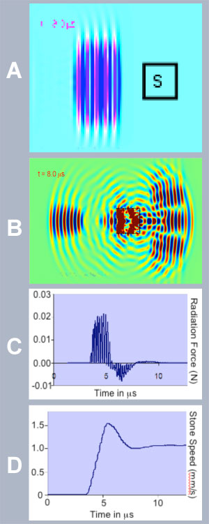
Although the applications are clear, the mechanism that causes twinkling is a complex mixture of factors. The ultrasound imager produces tiny motions in the stone and receives from the stone an echo that is generally stronger than that from tissue and contains reverberations from within the stone. These extra signals appear as if structures within the volume of the stone are moving in and out of the image. The confusion is further compounded by processing within the machine, which essentially amplifies the extra signals and variation in the collection of sequences of images. Our approach is to use numerical modeling of the echoes and reverberations, and compare these to the raw data collected by ultrasound images for stones in water, tissue, and patients. We then create our own images using specific algorithms that mimic the proprietary processing in the imaging systems. We can generally recreate what is shown on the imagers and detect patterns that are used to specifically image just stones and not motion. In our experience, the artifact reveals the stone in 100% of the animal studies and has performed reliably in an initial handful of human studies. Right: Modeling results suggest acoustic radiation force as a possible reason for the "twinkling artifact." A) Acoustic pressure distribution, when ultrasound diagnostic pulse is directed to a kidney stone (s), is scattered (B). The scattering gives rise to an oscillatory radiation force imparted on the stone (C). The stone is thus pushed and starts to move. The corresponding speed (D) is high enough to create a noticeable Doppler signal for small stones, less than 1 mm. Larger stones also can be pushed in the transverse direction, which may result in changing the ultrasound speckle pattern and also contribute to the artifact. |
 |
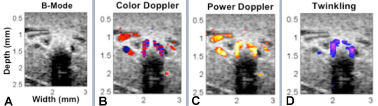 Above:Images generated on post-processing of raw data from the ultrasound imager: A) B-mode shows shadowing but little of the stone, B) and C) normal Doppler processing shows the artifact on the stone as well as blood flow, and D) new processing for the distinct signal of twinkling adds color only to the stone. |